最高のコレクション e coli structure diagram 194134-E coli structure diagram
Nanomaterials (NMs) including metals, metal oxides, and quantum dots are increasingly used for developing various materials of industrial use Conventional NM synthesis by chemical and physical methods requires rather harsh and environmentally hazardous conditions Here we report biosynthesis of various NMs by employing a recombinant Escherichia coli strain coexpressing metallothionein andEscherichia coli, when undergoing cellular division, is using a means of asexual reproduction because there is no transfer of genetic material;However, details of the molecular mechanism remain controversial Here we present the crystal structure at 22 A of MutS from Escherichia coli bound to a G x T mismatch
Plos One Structure Of Putrescine Aminotransferase From Escherichia Coli Provides Insights Into The Substrate Specificity Among Class Iii Aminotransferases
E coli structure diagram
E coli structure diagram-Draw along as I draw an Ecoli bacterium Marvel at my skills Go to the accompanying slideshow at http//wwwslidesharenet/jasondenys/ibbiology22prokaryADVERTISEMENTS In this article we will discuss about Escherichia Coli (E Coli) 1 Meaning of Escherichia Coli 2 Morphology and Staining of Escherichia Coli 3 Cultural Characteristics 4 Biochemical Reaction 5 Antigenic Structure 6 Toxin 7 Haemolysin 8 Infection E Coli Causes 9 Antigenic Typing 10 Laboratory Diagnosis 11 Treatment 12 Medical Importance Contents Meaning
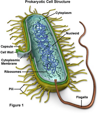


Molecular Expressions Cell Biology Bacteria Cell Structure
E coli is described as a Gramnegative bacterium This is because they stain negative using the Gram stain The Gram stain is a differential technique that is commonly used for the purposes of classifying bacteriaThe only structure thus far reported for TolR is of the periplasmic domain from Haemophilus influenzae in which N and Cterminal residues had been deleted (TolR(), Escherichia coli numbering) H influenzae TolR() is a symmetrical dimer with a large deep cleft at the dimer interfaceEscherichia coli (/ ˌ ɛ ʃ ə ˈ r ɪ k i ə ˈ k oʊ l aɪ /), also known as E coli (/ ˌ iː ˈ k oʊ l aɪ /), is a Gramnegative, facultative anaerobic, rodshaped, coliform bacterium of the genus Escherichia that is commonly found in the lower intestine of warmblooded organisms (endotherms) Most E coli strains are harmless, but some serotypes (EPEC, ETEC etc) can cause serious food
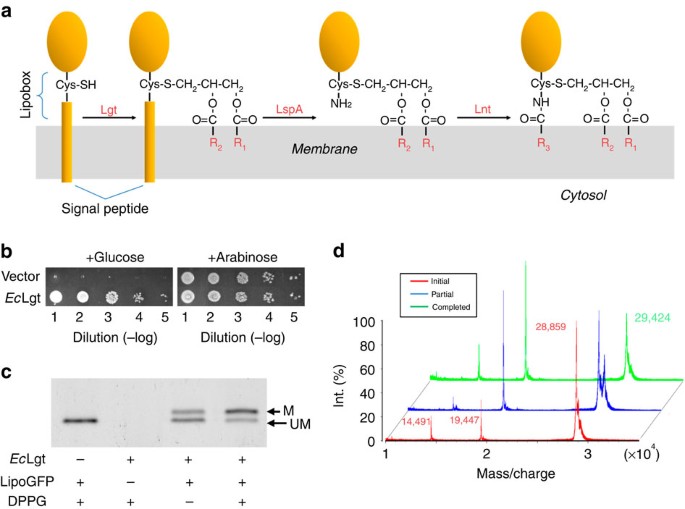
Escherichia coli (abbreviated as E coli) are bacteria found in the environment, foods, and intestines of people and animalsE coli are a large and diverse group of bacteria Although most strains of E coli are harmless, others can make you sick Some kinds of E coli can cause diarrhea, while others cause urinary tract infections, respiratory illness and pneumonia, and other illnessesIn the form1 Ec Lgt structure with a palmitic acid and an OG molecule, the guanidine group of R143 in the signature motif is found to bind with the carboxyl headgroup of palmitic acid (Fig 5a)Draw and label a diagram of the ultrastructure of Escherichia coli (E coli ) as an example of a prokaryote Annotate the diagram of prokaryotic cell with the functions of each named structure Identify structures in electron micrographs of E coli State that prokaryotic cells divide by binary fission EUKARYOTIC CELLS
The structure of LsrK was not known until 18 when the crystal structure of E coli LsrK (open form) was released including a modulator protein called HPr (PDB ID 5YA0, 5YA1, and 5Y) (19) Structurally, LsrK belongs to the FGGY carbohydrate kinases (sugar) () consisting of two domains Domain I (of Nterminal residues 13–260) and DomainThe structure of LsrK was not known until 18 when the crystal structure of E coli LsrK (open form) was released including a modulator protein called HPr (PDB ID 5YA0, 5YA1, and 5Y) (19) Structurally, LsrK belongs to the FGGY carbohydrate kinases (sugar) () consisting of two domains Domain I (of Nterminal residues 13–260) and DomainThe only structure thus far reported for TolR is of the periplasmic domain from Haemophilus influenzae in which N and Cterminal residues had been deleted (TolR(), Escherichia coli numbering) H influenzae TolR() is a symmetrical dimer with a large deep cleft at the dimer interface


Plos One Structure Of Putrescine Aminotransferase From Escherichia Coli Provides Insights Into The Substrate Specificity Among Class Iii Aminotransferases
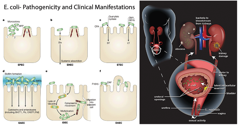


Escherichia Coli E Coli An Overview Microbe Notes
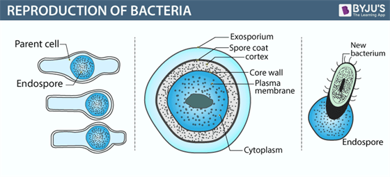
E coli is a type of bacteria that normally live in the intestines of people and animals However, some types of E coli , particularly E coli O157H7, can cause intestinal infectionInstead, their genetic material floats uncovered, localized to a region called the nucleoidWhen there is a favourable condition, Ecoli or Escherichia coli produces about 2 million bacteria every 7 hours To know more about bacteria, its definition, the structure of bacteria, bacteria diagram, classification of bacteria, and reproduction in bacteria keep visiting BYJU'S website or download BYJU'S app for further reference
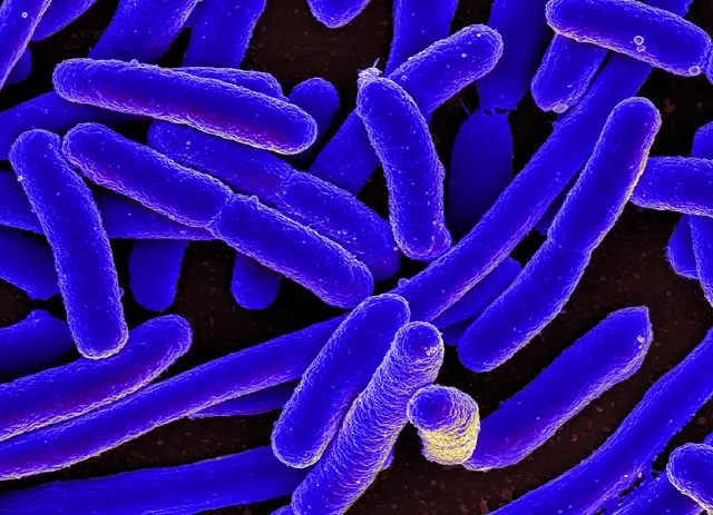


E Coli Under The Microscope Types Techniques Gram Stain Hanging Drop Method
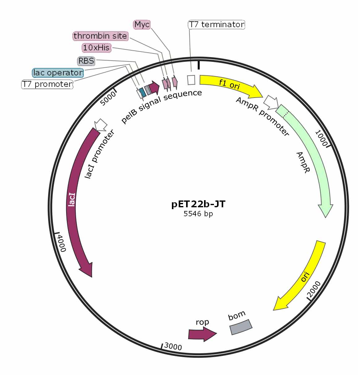


How To Choose A Suitable Expression Vector
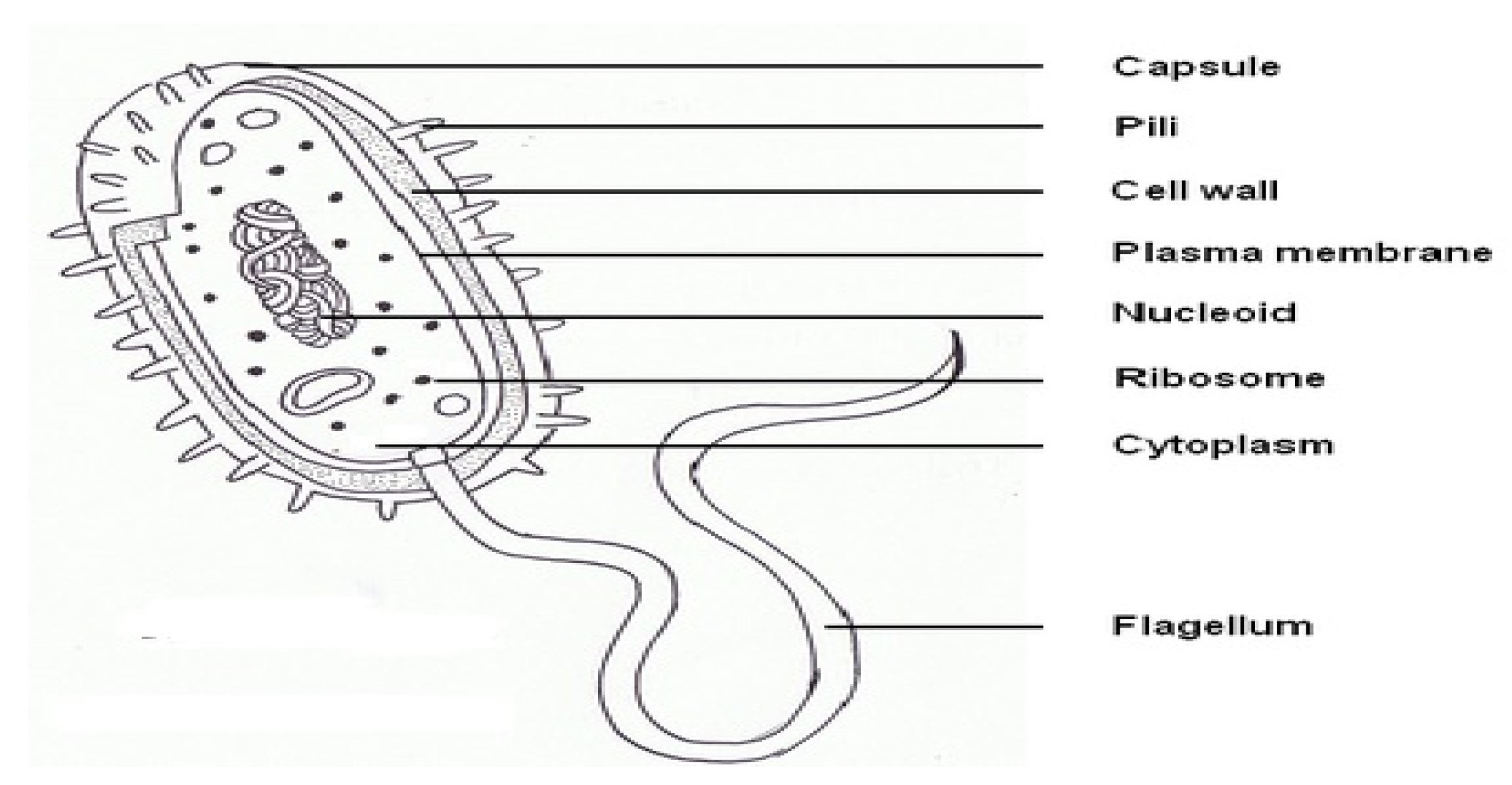
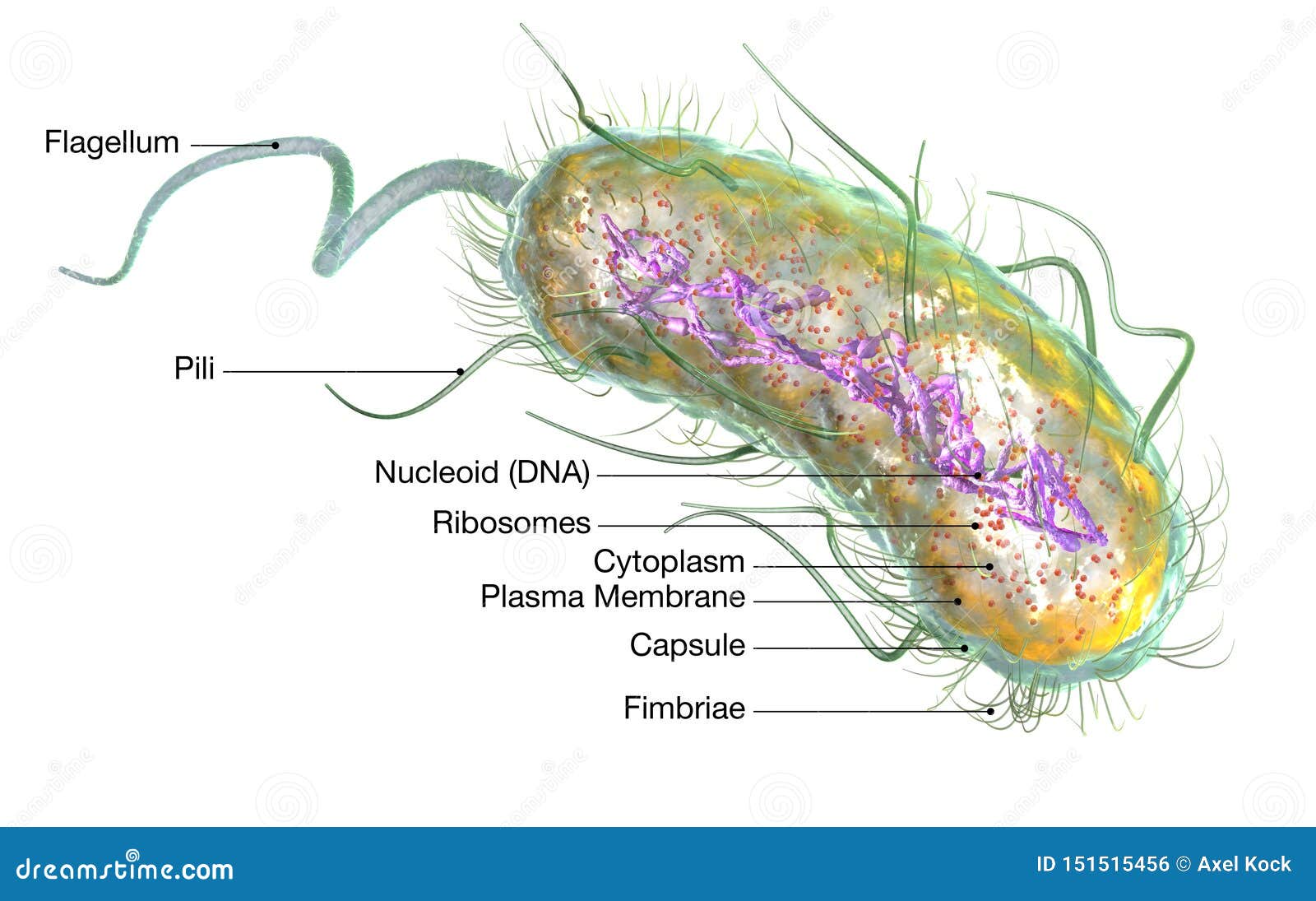
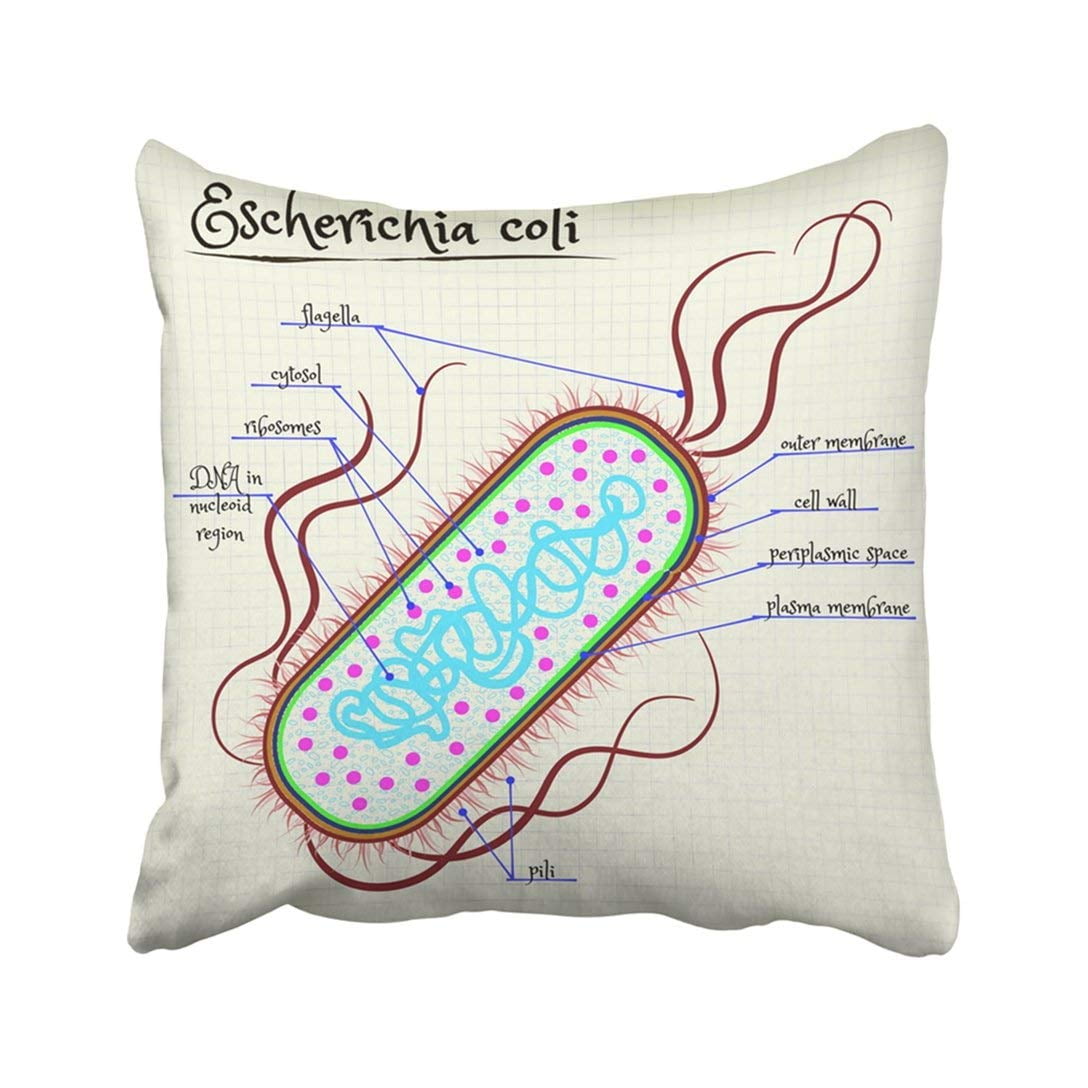
E coli are rodshaped bacterium that has an outer membrane consisting of lipopolysaccharides, inner cytoplasmic membrane, peptidoglycan layer, and an inner, cytoplasmic membrane Cell wall this structure is made of a thick layer of protein and sugar that prevents the cell from bursting Plasma membrane this structure is made of lipids and proteins and is used to control the movement of molecules in and out the cellIn the form1 Ec Lgt structure with a palmitic acid and an OG molecule, the guanidine group of R143 in the signature motif is found to bind with the carboxyl headgroup of palmitic acid (Fig 5a)In Escherichia coli, the peptidoglycan crosslinking reaction to form the cell wall is primarily carried out by penicillinbinding proteins that catalyse D,Dtranspeptidase activity However, an alternate crosslinking mechanism involving the L,Dtranspeptidase YcbB can lead to bypass of D,Dtranspeptidation and betalactam resistance


Draw And Label A Diagram Of The Ultrastructure Of Escherichia Coli E Ppt Download


Electron Transport Chain Of Bacteria With Diagram
The bacterium is merely making an exact copy of itself (watch out for the a ttack of the Clones!) This is the most prevalent form of reproduction for E coliEscherichia coli is commonly found in the normal microflora in the human gastrointestinal tract and is intricately involved in the lives of humansThis bacterium can be grown readily, and its genetics are easily manipulated in the laboratory, making it a common workhorse and one of the beststudied prokaryotic model organismsWhen there is a favourable condition, Ecoli or Escherichia coli produces about 2 million bacteria every 7 hours To know more about bacteria, its definition, the structure of bacteria, bacteria diagram, classification of bacteria, and reproduction in bacteria keep visiting BYJU'S website or download BYJU'S app for further reference


E Coli Medical Images For Power Point


Structure Of The Escherichia Coli Rna Polymerase A Subunit Amino Terminal Domain Science
NMR studies of the E coli 5S rRNA fragment containing loop E have shown once again that singlestranded regions in RNA molecules may have rather well ordered structure In particular, the loop E appears to resemble a double helix but formed by nonWatsonCrick base pairs such as GG, GA, reversed Hoogsteen AU, and GU stabilized by a single hydrogen bond (Fig 67)Overall structure and topology of E coliPBP1b (A) The crystal structure of PBP1b is represented as a ribbon diagram The TM, UB2H, TG, and TP domains are color coded in cyan, yellow, red, and blue, respectively Moenomycin is represented as van derThe crystal structure of E coli MgsA revealed three apparent structural domains in the protein as follows Nterminal ATPbinding (residues ∼22–165) and adjacent helical lid domains (residues ∼166–247 and a short Nterminal helix (14–21)) that are conserved among AAA proteins and a Cterminal tetramerization domain (residues ∼251–446) (Fig 1 A)


Crystal Structures Of The E Coli Transcription Initiation Complexes With A Complete Bubble Sciencedirect
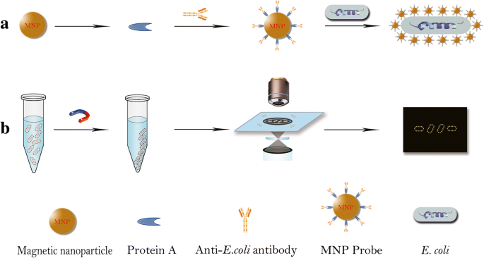


Ultrasensitive And Rapid Count Of Escherichia Coli Using Magnetic Nanoparticle Probe Under Dark Field Microscope Bmc Microbiology Full Text
MurNAc is unique to bacterial cell walls, as is Dglu, DAP and Dala The muramic acid subunit of E coli is shown in Figure 16 below Figure 16 The structure of the muramic acid subunit of the peptidoglycan of Escherichia coli This is the type of murein found in most Gramnegative bacteriaE Coli reproduce by binary fission and conjugation ("the transfer of genetic material through a sex pilus") The most prevalent reproduction for E Coli is asexual reproduction which takes place when the E Coli is undergoing binary fission This type of reproduction begins with the replication of one DNA moleculeCauses Only a few strains of E coli trigger diarrhea The E coli O157H7 strain belongs to a group of E coli that produces a powerful toxin that damages the lining of the small intestine This can cause bloody diarrhea You develop an E coli infection when you ingest this strain of bacteria Unlike many other diseasecausing bacteria, E coli can cause an infection even if you ingest


Bacterial Flagella Structure Importance And Examples Of Flagellated Bacteria Learn Microbiology Online


Draw The Structure Of E Coli Label Those Structures Which Differentiate Them From Eukaryotic Cells Biology Cell The Unit Of Life Meritnation Com
E coli, (Escherichia coli), species of bacterium that normally inhabits the stomach and intestines When E coli is consumed in contaminated water, milk, or food or is transmitted through the bite of a fly or other insect, it can cause gastrointestinal illness Mutations can lead to strains that cause diarrhea by giving off toxins, invading the intestinal lining, or sticking to the intestinalE coli is a Gramnegative rodshaped bacteria, which possesses adhesive fimbriae and a cell wall that consists of an outer membrane containing lipopolysaccharides, a periplasmic space with a peptidoglycan layer, and an inner, cytoplasmic membraneThe crystal structure at 39 angstroms resolution demonstrates that Escherichia coli MscS folds as a membranespanning heptamer with a large cytoplasmic region Each subunit contains three


Phylogenetic Background And Habitat Drive The Genetic Diversification Of Escherichia Coli Biorxiv
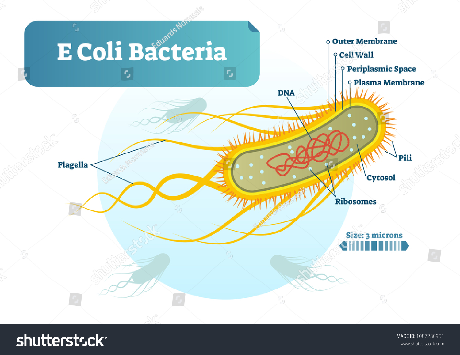


E Coli Bacteria Micro Biological Vector Stock Vector Royalty Free
Structure And Function Of A Ecoli Bacteria E coli is a rodshaped bacterium The peptidoglycan cell wall is thin and multilayered There is a thin peptidoglycan layer placed between the inner cytoplasmic membrane, and the outer membrane The outer membrane is surrounded by the capsule layer which is composed of polysaccharides The flagellum of E coli consists of three distinct parts the filament, the hook and the basal body all of which are made up of different types of proteinsOverview Escherichia coli (E coli) is a bacterium that is commonly found in the gut of humans and warmblooded animalsMost strains of E coli are harmless Some strains however, such as Shiga toxinproducing E coli (STEC), can cause severe foodborne disease It is transmitted to humans primarily through consumption of contaminated foods, such as raw or undercooked ground meat products, rawE coli is a common bacteria that lives in the lower gastrointestinal tract of humans and animals It also can be isolated from water and soil Although most strains are harmless, some strains of E coli are capable of producing powerful toxins that can cause severe illness


1 2 Prokaryotic Cells A Biology


The Ding Protein From Escherichia Coli Is A Structure Specific Helicase Journal Of Biological Chemistry
It is a tough and rigid structure of peptidoglycan with accessory specific materials (eg LPS, teichoic acid etc) surrounding the bacterium like a shell and lies external to the cytoplasmic membrane It is 1025 nm in thickness It gives shape to the cell Nucleus The single circular doublestranded chromosome is the bacterial genomeRecognition of a DNA mismatch by a dimeric MutS protein initiates a cascade of reactions and results in repair of the newly synthesized strand;Escherichia coli (E coli) bacteria live in the intestines of people and animals, and are key to a healthy intestinal tract Most E coli strains are harmless, but some can cause diarrhea through contact with contaminated food or water while other strains can cause urinary tract infections, respiratory illness and pneumonia


Electron Microscopy Images As Well As A Sketch Of The E Coli And The Download Scientific Diagram


Prokaryotic Cell E Coli Structure And Definition Diagram Quizlet
Illustration about Escherichia coli bacteria structure diagram vector illustration E coli info graphic Illustration of medical, diagram, healthyEnteroaggregative E coli (EAEC) are a heterogeneous collection of strains characterized by their autoagglutination in a "stackedbrick" arrangement over the epithelium of the small intestine and, in some cases, the colon That is, it is so named because it adheres to HEP2 cells in a distinct pattern, layering of the bacteria aggregated in a stackedbrick fashionMost strains of E coli are rodshaped and measure about μm long and 0210 μm in diameter They typically have a cell volume of 0607 μm, most of which is filled by the cytoplasm Since it is a prokaryote, E coli don't have nuclei;


Diagram E Coli Structure Hd Png Download 2508x1747 Png Dlf Pt


Structure And Function Of Bacterial Cells
Causes Only a few strains of E coli trigger diarrhea The E coli O157H7 strain belongs to a group of E coli that produces a powerful toxin that damages the lining of the small intestine This can cause bloody diarrhea You develop an E coli infection when you ingest this strain of bacteria Unlike many other diseasecausing bacteria, E coli can cause an infection even if you ingestThe growth of an E coli colony over time Credit WikiCommons CCBY SA 40 The Role Of E coli In The Human Body And Disease Most strains of E coli are actually completely harmless to humans In fact, colonies of E coli form a natural part of the microbiome of the mammalian gut In mammals, E coli facilitates the absorption of iron via the production of enterobactin, a siderophore (GreekBacteria species E coli and S aureus under the microscope with different magnifications Bacteria are among the smallest, simplest and most ancient living


Q Tbn And9gctffwwm42evo5c26svvkxccrastvfjru302ru 63nxjqythb6cl Usqp Cau
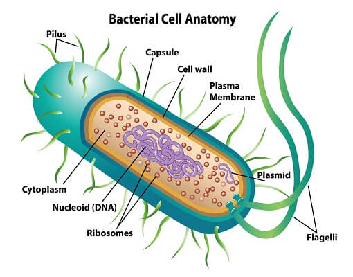


Prokaryotic Cell Definition Examples Structure Biology Dictionary
E coli, (Escherichia coli), species of bacterium that normally inhabits the stomach and intestines When E coli is consumed in contaminated water, milk, or food or is transmitted through the bite of a fly or other insect, it can cause gastrointestinal illness Mutations can lead to strains that cause diarrhea by giving off toxins, invading the intestinal lining, or sticking to the intestinal_____ is named as such because it is an EColi but it does not cause the pink colonies on the MacConkey plate Upper level primate pathogen Look at the lacoperon (there is a bacteriophage there that blocks the lactose fermentation) to determine what it isIt is a tough and rigid structure of peptidoglycan with accessory specific materials (eg LPS, teichoic acid etc) surrounding the bacterium like a shell and lies external to the cytoplasmic membrane It is 1025 nm in thickness It gives shape to the cell Nucleus The single circular doublestranded chromosome is the bacterial genome
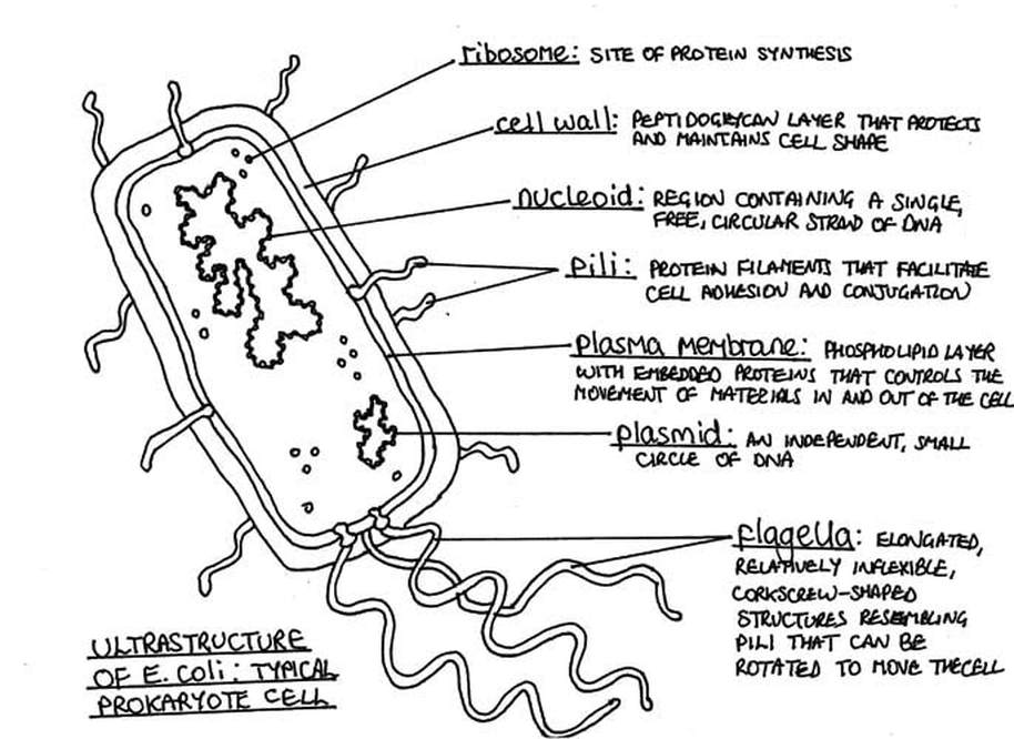


Ib Hl 1 2 S1


Structural Bases For F Plasmid Conjugation And F Pilus Biogenesis In Escherichia Coli Pnas
E Coli reproduce by binary fission and conjugation ("the transfer of genetic material through a sex pilus") The most prevalent reproduction for E Coli is asexual reproduction which takes place when the E Coli is undergoing binary fission This type of reproduction begins with the replication of one DNA moleculeE coli is a common bacteria that lives in the lower gastrointestinal tract of humans and animals It also can be isolated from water and soil Although most strains are harmless, some strains of E coli are capable of producing powerful toxins that can cause severe illnessRibosephosphate pyrophosphokinase (EC 2761) is an enzyme that catalyzes the ATPdependent conversion of ribose5phosphate to phosphoribosyl pyrophosphate The reaction product is a key precursor for the biosynthesis of purine and pyrimidine nucleotides We report the 22 Å crystal structure of the E coli ribosephosphate pyrophosphobinase (EcKPRS)
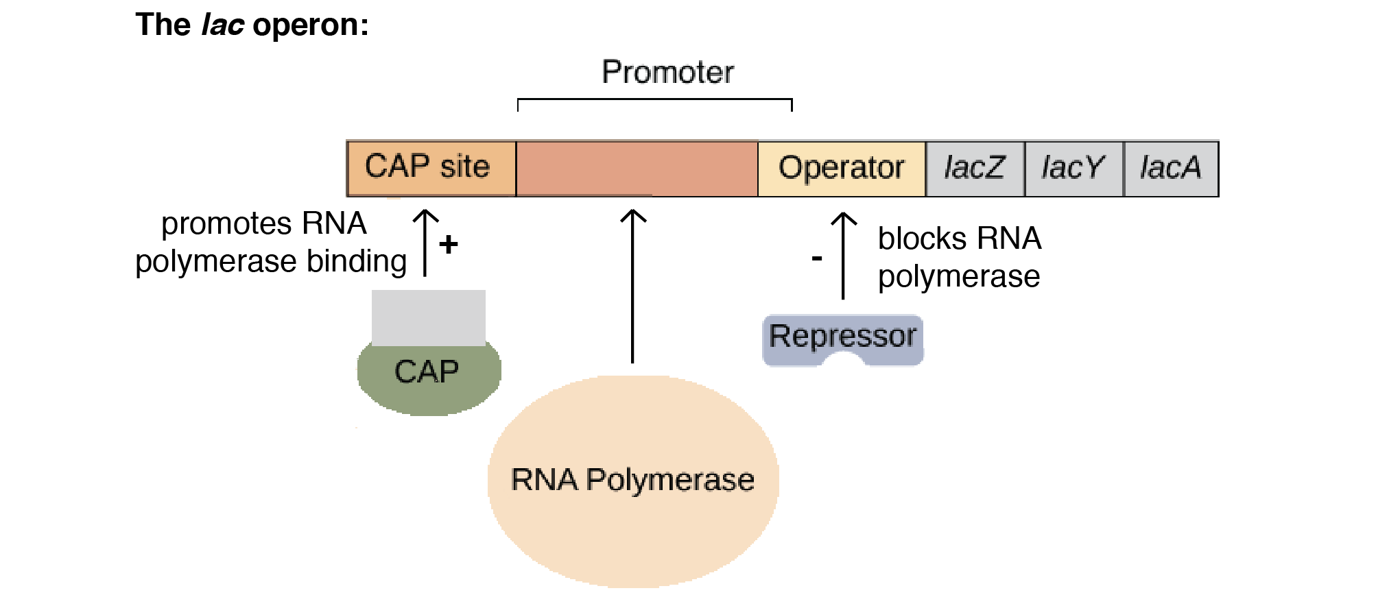


The Lac Operon Article Khan Academy


Escherichia Coli Wikipedia
Enteroaggregative E coli (EAEC) are a heterogeneous collection of strains characterized by their autoagglutination in a "stackedbrick" arrangement over the epithelium of the small intestine and, in some cases, the colon That is, it is so named because it adheres to HEP2 cells in a distinct pattern, layering of the bacteria aggregated in a stackedbrick fashionEscherichia coli (abbreviated as E coli) are bacteria found in the environment, foods, and intestines of people and animalsE coli are a large and diverse group of bacteria Although most strains of E coli are harmless, others can make you sick Some kinds of E coli can cause diarrhea, while others cause urinary tract infections, respiratory illness and pneumonia, and other illnessesOverall structure and topology of E coliPBP1b (A) The crystal structure of PBP1b is represented as a ribbon diagram The TM, UB2H, TG, and TP domains are color coded in cyan, yellow, red, and blue, respectively Moenomycin is represented as van der


Crystal Structure Of Udp N Acetylmuramoyl L Alanyl D Glutamate Meso Diaminopimelate Ligase From Escherichia Coli Journal Of Biological Chemistry


Q Tbn And9gcsaiyw62wcbrvh1nz2sr H2o5hyidohj9bi9jyz223ejd Onqw Usqp Cau
E coli is Gramnegative straight rod, 13 µ x 0407 µ, arranged singly or in pairs (Fig 281) It is motile by peritrichous flagellae, though some strains are nonmotile Spores are not formed Capsules and fimbriae are found in some strainsNonpathogenic Escherichia coli strain Nissle 1917 (O6K5H1) is used as a probiotic agent in medicine, mainly for the treatment of various gastroenterological diseases To gain insight on the genetic level into its properties of colonization and commensalism, this strain's genome structure has been analyzed by three approaches (i) sequence context screening of tRNA genes as a potential


Chromatinization Of Escherichia Coli With Archaeal Histones Elife


Onlinelibrary Wiley Com Doi Pdf 10 1111 J 1365 2958 05 X


Physical And Functional Interactions Between Recq And Ssb Crystal Download Scientific Diagram
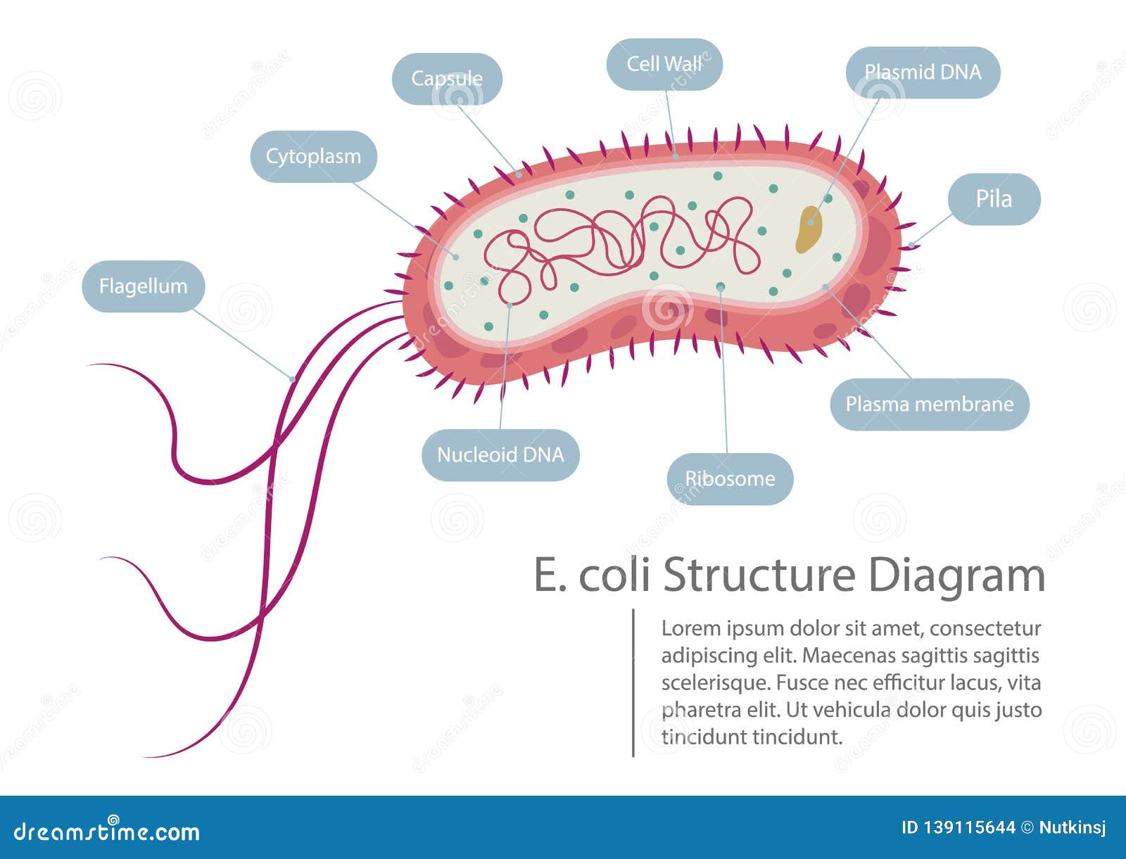


Escherichia Coli Structure Diagram Stock Vector Illustration Of Medical Diagram


E Coli Bacteria Micro Biological Vector Illustration Cross Section Labeled Diagram Medical Research Information Poster Clip Art K Fotosearch


Prokaryotic Cells Bioninja


Pin On Health And Medicine Illustrated
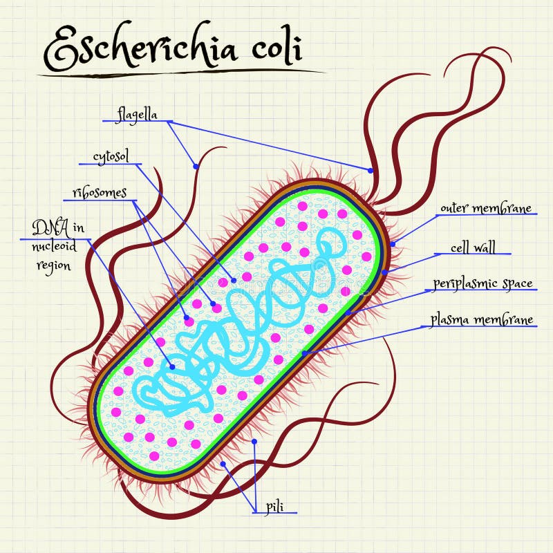


The Structure Of Escherichia Coli Stock Vector Illustration Of Ribosomes Colorful


Qs 21 The Drawing Below Shows The Ultrastructure Of E Coli Ppt Video Online Download


The Cryo Em Structure Of A Translation Initiation Complex From Escherichia Coli Cell
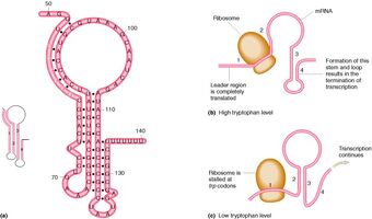


Escherichia Coli Trp Operon Mmg 233 14 Genetics Genomics Wiki Fandom


Applied Sciences Free Full Text Photocatalytic Inactivation As A Method Of Elimination Of E Coli From Drinking Water Html


Escherichia Coli Adhesins And Harboring Motile Structures Download Scientific Diagram


Ligand And Structure Based Approaches Of Escherichia Coli Fabi Inhibition By Triclosan Derivatives From Chemical Similarity To Protein Dynamics Influence Chemmedchem X Mol


Rcsb Pdb 6c9y Cryo Em Structure Of E Coli Rnap Sigma70 Holoenzyme


Solved 4 Ill Biochemical Structures Diagr 1 Diagram The Chegg Com


Structure Of The E Coli Hbp Passenger And Its Ac Domain A Ribbon Download Scientific Diagram


Vector Clipart E Coli Bacteria Micro Biological Vector Illustration Cross Section Labeled Diagram Medical Research Information Poster Vector Illustration Gg Gograph


E Coli Ribosome Structure


Molecular Expressions Cell Biology Bacteria Cell Structure


Lac Operon Definition Structure Inducers Diagram


2 2 Prokaryotic Cells Contents 1 Assessment Statements 2 Lecture Notes 3 Slide Show 4 On Line Resources 5 Reference Sites Assessment Statements Assessment Statement Obj Teacher S Notes 2 2 1 Draw And Label A Diagram Of The Ultrastructure Of


The Structure Of Escherichia Coli Stock Illustration Download Image Now Istock
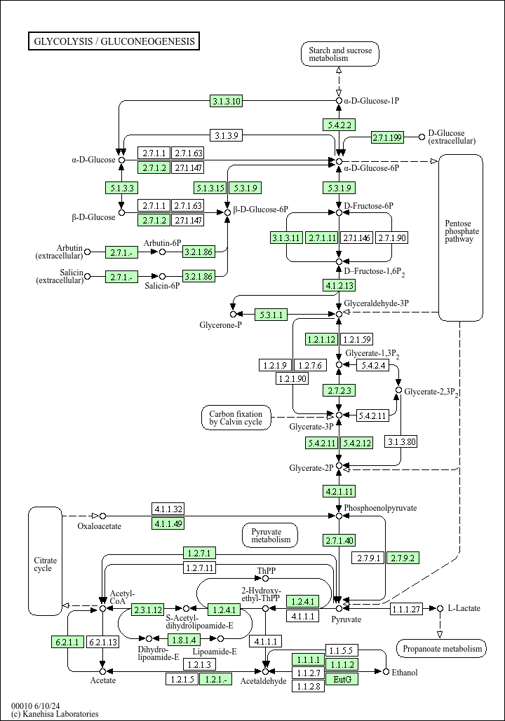


Kegg Pathway Glycolysis Gluconeogenesis Escherichia Coli K 12 Mg1655
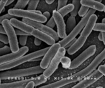


Escherichia Coli Microbewiki


A Secondary Structure Diagram Of E Coli P Rna Helices Are Download Scientific Diagram


3
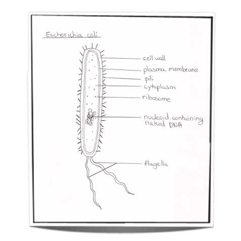


Ib Biology Notes 2 2 Prokaryotic Cells


Crystal Structure Of Escherichia Coli Pdxa An Enzyme Involved In The Pyridoxal Phosphate Biosynthesis Pathway Journal Of Biological Chemistry


E Coli Annotation Clip Art Library


Q Tbn And9gcqkye60ou Johpr02n Mbv1fferrjpdh Lnct7ymdf5qhyia1ld Usqp Cau
/shigella-bacteria--illustration-758308491-5a02252f9e9427003c1759be.jpg)


Prokaryotic Cells Structure Function And Definition
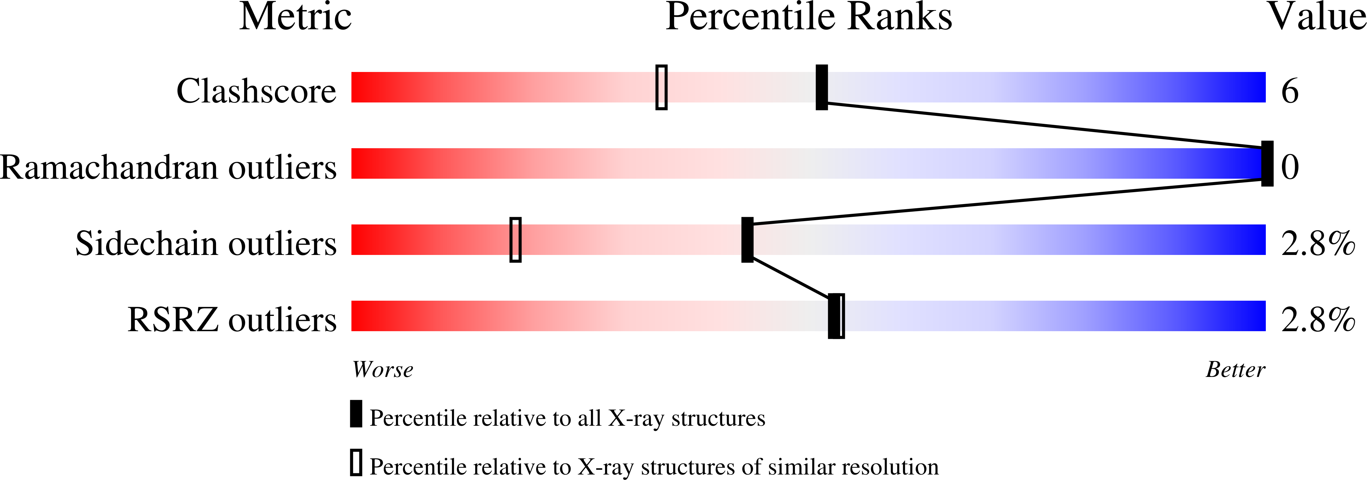


Rcsb Pdb 1t3m Structure Of The Isoaspartyl Peptidase With L Asparaginase Activity From E Coli
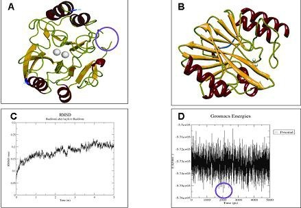


A Crystallized 3d Structure Of Blandm 1 In E Coli Ma Open I


Fine Scale Structure Analysis Shows Epidemic Patterns Of Clonal Complex 95 A Cosmopolitan Escherichia Coli Lineage Responsible For Extraintestinal Infection Msphere
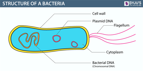


Bacteria Definition Structure Diagram Classification


V5nj3lqzqc0ksm
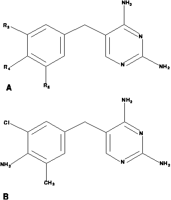


Discovering Rules For The Inhibition Of E Coli Dihydrofolate Reductase H2
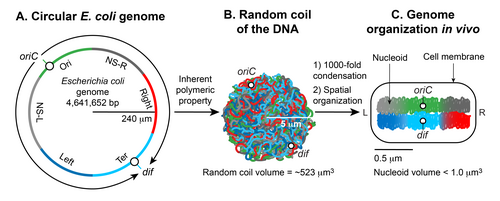


Nucleoid Wikipedia


Crystal Structure Of E Coli Lipoprotein Diacylglyceryl Transferase Nature Communications


X Ray Crystal Structure Of The E Coli Ompf Porin A Ribbon Diagram Of The Monomer


E Coli Cell Diagram Page 5 Line 17qq Com


Pathogenic Escherichia Coli Nature Reviews Microbiology


The Structure Of The Ubiquinol Oxidase From Escherichia Coli And Its Ubiquinone Binding Site Nature Structural Molecular Biology


E Coli Diagram Drone Fest
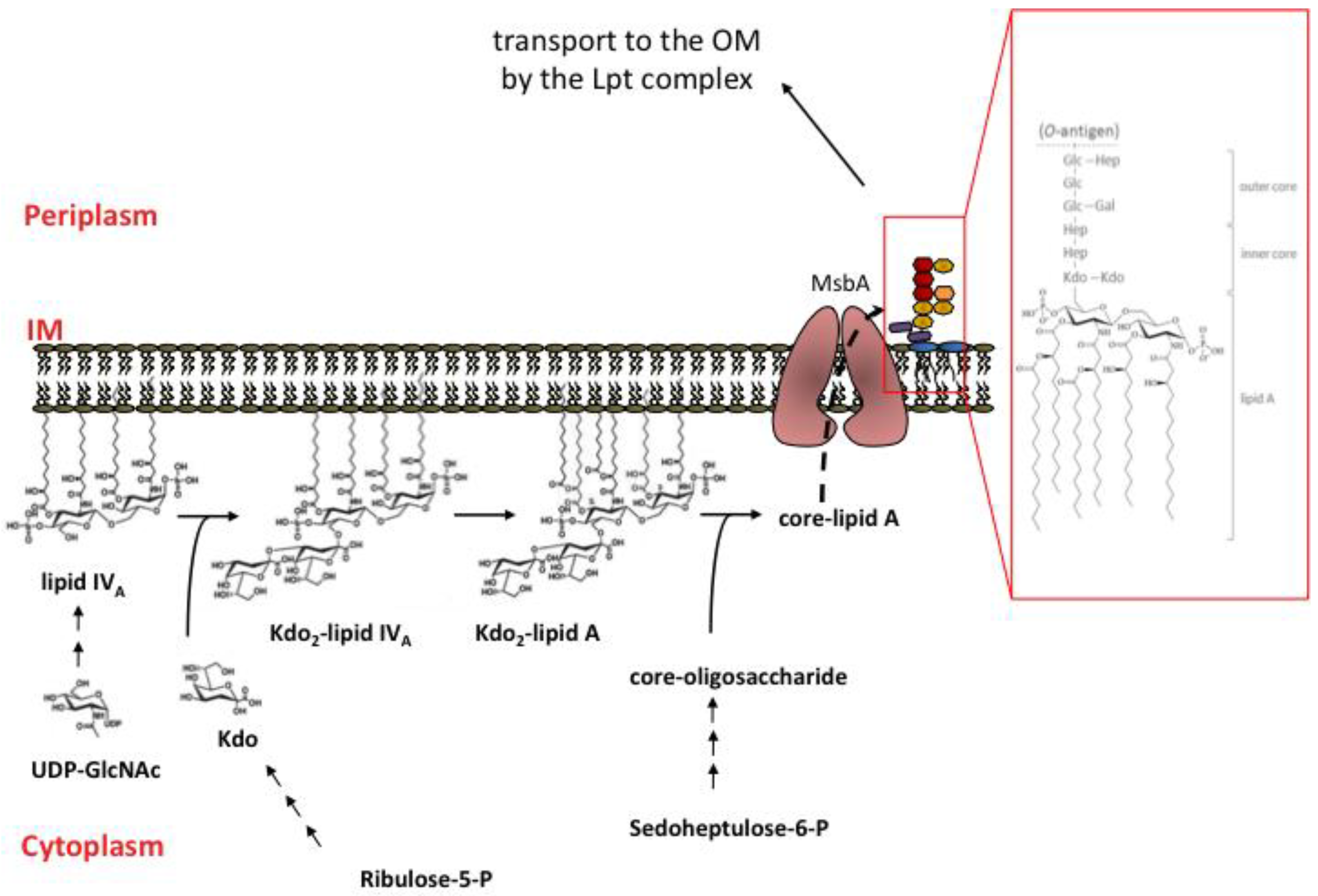


Marine Drugs Free Full Text The Lipopolysaccharide Export Pathway In Escherichia Coli Structure Organization And Regulated Assembly Of The Lpt Machinery Html
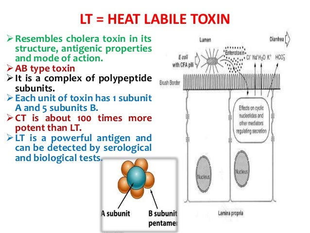


Genus Escherichia Coli


What Is Bacterial Endotoxin Wako Lal System


Characterization Of The Two Protein Complex In Escherichia Coli Responsible For Lipopolysaccharide Assembly At The Outer Membrane Pnas


Diversity Of Lsu Mt Rrnas The Secondary Structure Diagram Of Download Scientific Diagram


Www Tustin K12 Ca Us Uploaded Schools Foothill Documents Ss Ib Bio Hl Pdf


Worldwide Population Structure Of Escherichia Coli Reveals Two Major Subspecies Biorxiv


2 2 Prokaryotic Cells Bioninja


Multiscale Structuring Of The E Coli Chromosome By Nucleoid Associated And Condensin Proteins Sciencedirect
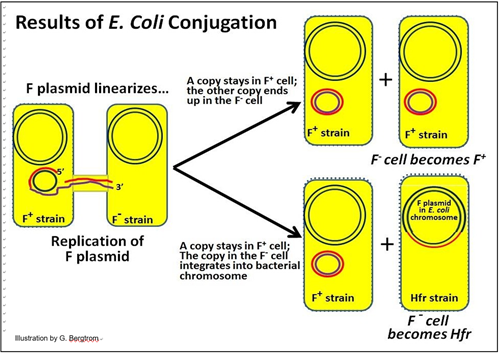


8 5 Structure And Organization Of Dna In Bacteria Biology Libretexts
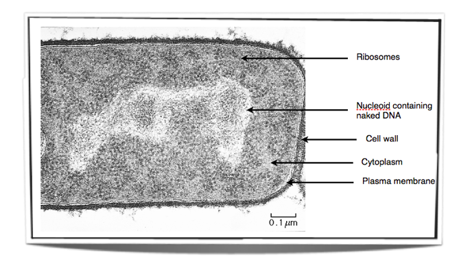


Ib Biology Notes 2 2 Prokaryotic Cells


Human Point Mutations Mapped Onto The E Coli Ssadh Structure


E Coli Bacteria Micro Biological Vector Illustration Cross Section Labeled Diagram Medical Research Information Poster Canvas Print Barewalls Posters Prints Bwc


Rcsb Pdb 2gs4 The Crystal Structure Of The E Coli Stress Protein Ycif


Pdf Structure And Regulation Of Escherichia Coli Isocitrate Dehydrogenase Semantic Scholar


Entamoeba Coli Wikipedia


2 2 1 Draw And Label A Diagram Of The Ultrastructure Of Ecoli As An Example Of A Prokaryote Youtube


Escherichia Coli Wikipedia


Diagram Of Prokaryotic Cell Teacher Example


Escherichia Coli Bacteria E Coli Medically Accurate 3d Illustration Labeled Stock Illustration Illustration Of Biota Biology


E Coli Chemotaxis


Replication Termination In Escherichia Coli Structure And Antihelicase Activity Of The Tus Ter Complex Microbiology And Molecular Biology Reviews


2 2 Prokaryotic Cells Bioninja


Bpbop Colorful Chart Drawing Of The Structure Escherichia Coli Color Diagram Dna Education Pillowcase Throw Pillow Cover Case 16x16 Inches Walmart Com Walmart Com


Crystal Structure Of Escherichia Coli Cyay Protein Reveals A Previously Unidentified Fold For The Evolutionarily Conserved Frataxin Family Pnas


Pin On Science


Escherichia Coli


Architecture Of The Escherichia Coli Nucleoid


Bacteria Definition Structure Diagram Classification


Flagellum Structure Diagram Quizlet
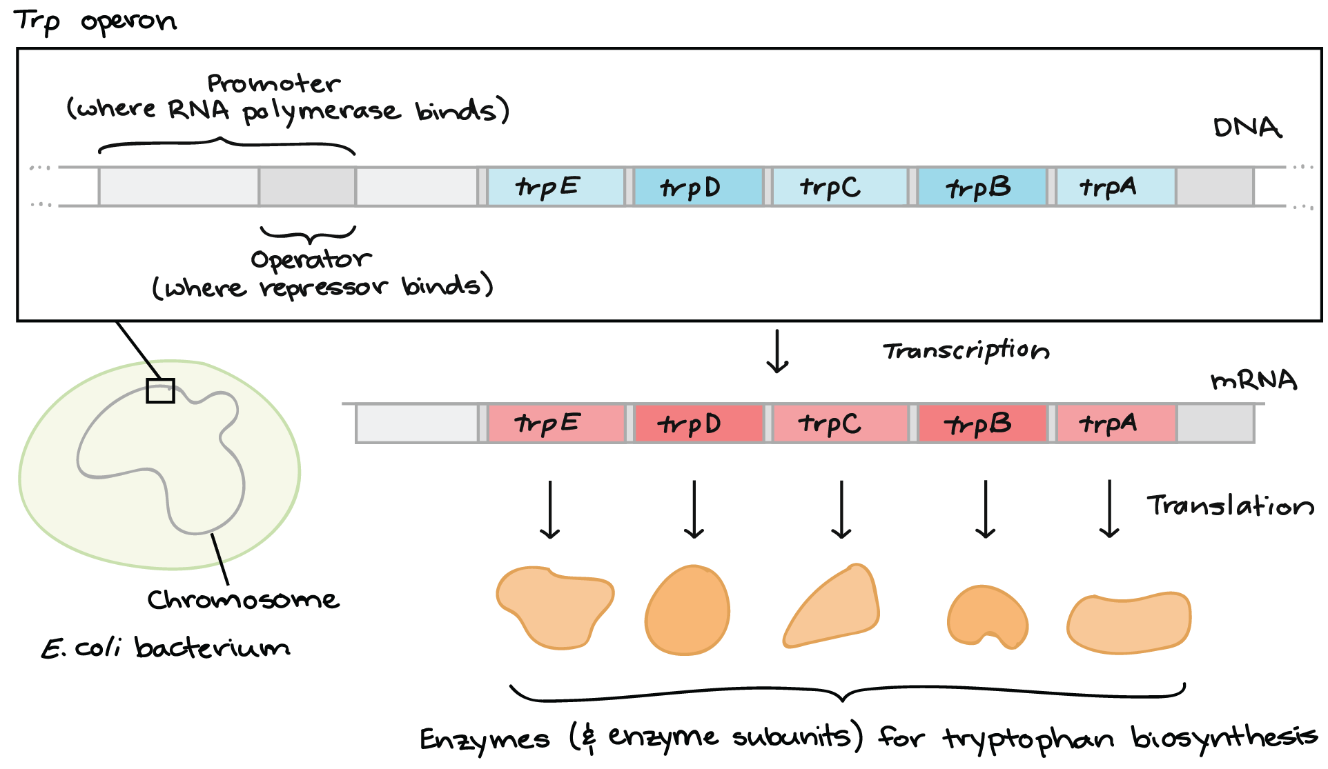


The Trp Operon Article Khan Academy


コメント
コメントを投稿